Why teaching AI about symmetry could make prenatal diagnosis more reliable
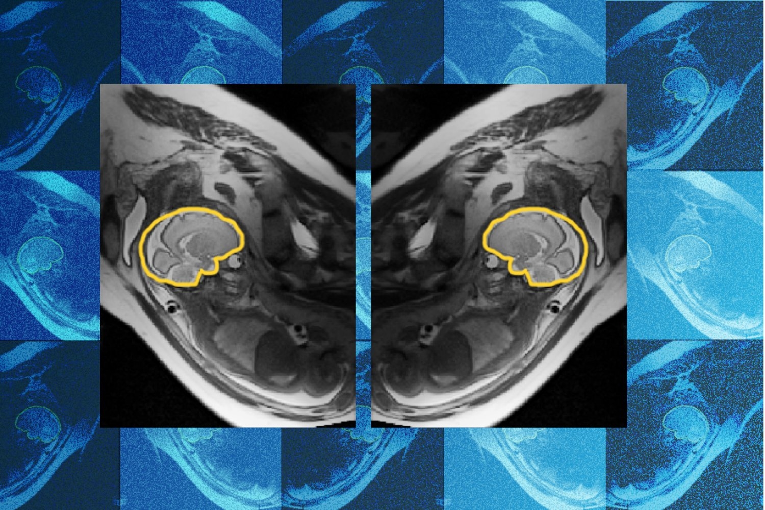
Researchers at MIT, Boston Children's Hospital and the French research institute Inria have developed and trained a deep learning model called E(3)-Pose that tracks a fetal head while it moves during a magnetic resonance imaging scan, with the aim of producing a high-resolution image a radiologist can actually read. A paper describing the work was presented at the 2026 European Conference on Computer Vision. The first author is Ramya Muthukrishnan, an MIT PhD student in the lab of Polina Golland, a professor of electrical engineering and computer science who is a principal investigator at the MIT Abdul Latif Jameel Clinic for Machine Learning in Health and at MIT CSAIL.
The problem the model addresses is mechanical. A fetal MRI is built from a series of 2D images, and those planes are stacked to form one high-resolution 3D picture of the head. If the fetus shifts mid-scan, each slice can be captured from a different angle, leaving gaps in the final volume and an unusual slice orientation. A physician reviewing such a scan may then miss a pathology or diagnose one incorrectly. Muthukrishnan frames the stakes in terms of resolution: fine details require a good volumetric picture, and coverage gaps can hide critical brain regions at exactly the moment a clinician is looking for signs of abnormal development.
Clinically, the motivation is concrete. A pregnant patient may spend an hour in the scanner and quit partway through because of discomfort, leaving the radiologist without a complete, high-quality study. Muthukrishnan describes shadowing a radiologist performing fetal MRI at Boston Children's and seeing that frustration on both sides of the exam, which she says helped set her PhD direction.
What distinguishes E(3)-Pose, per the researchers, is where the physics lives. Instead of asking the network to infer physical structure purely from training data, the group embedded a known property of the anatomy — the left-right symmetry of the fetal head — directly into the model architecture. That prior makes it easier for the network to pin down the brain's orientation as it moves. In machine learning terms this belongs to a family of geometric priors: symmetry and equivariance constraints baked into a model rather than learned. Here the payoff is plausible because the constraint is true of the object being imaged; a symmetry assumption that did not hold would bias the estimate instead of steadying it.
Getting the model from a GPU onto real hardware was its own ordeal. Muthukrishnan, whose background was in computer science rather than physics or MRI physics, says she was unprepared for how often she crashed the scanner while implementing the method on the machine, and that deploying to hardware is a different beast from training on data. Co-author Borjan Gagoski, an assistant professor of radiology at Harvard Medical School and a researcher at Boston Children's, developed a sequence that acquires additional MRI scans to support training and helped troubleshoot the scanner when it failed. Co-author Aryn Lee, a research assistant at Boston Children's, recruited pregnant volunteers and assisted with acquiring each scan. Senior authors included Elfar Adalsteinsson, MIT professor of electrical engineering and computer science; Ellen Grant, the Käthe Beutler Professor of Pediatrics and professor of radiology at Harvard Medical School; and Golland. Benjamin Billot, a researcher at Inria and a former postdoc in Golland's group, helped Muthukrishnan get started with an earlier method he had developed.
Golland characterizes the contribution as combining geometric ideas such as equivariance and symmetry with deep learning to change how scanners acquire images, with an emphasis on how the work models image acquisition itself. Grant, who directs the Fetal Neonatal Neuroimaging and Developmental Science Center at Boston Children's, says the work will automate fetal brain MRI, replacing a slow manual process with faster and more comfortable scans for mothers while giving radiologists higher-quality images to interpret. Those are forward-looking characterizations from the project's senior authors rather than reported clinical results; the announcement cites no accuracy figures, patient counts or comparison against existing motion-correction pipelines, so the size of the gain is not yet something a reader can weigh.
Scope is also bounded. The model is primarily trained on third-trimester fetuses. Muthukrishnan says the next goal is second-trimester and younger cases, which are harder because those fetuses are small and move more in the available space of the uterus. She argues they matter more, since earlier detection of brain abnormalities makes intervention easier. That framing points to a likely development pattern for this kind of medical imaging model: performance tuned on later-gestation anatomy, then stretched toward the smaller, more mobile cases where the clinical value is highest and the imaging problem is hardest.
Beyond the womb, the group suggests the approach could help image other anatomy that is difficult to capture and even support autonomous driving, where limited sensors must make sense of noisy 3D data to track objects in three dimensions. That is a broad claim, and the shared thread — estimating pose from partial, motion-corrupted observations using known geometric structure — is the part most likely to transfer.
The work was supported by the MIT Jameel Clinic, the MIT Takeda Program, the Chou Family Transformative Research Fund, the National Institute of Biomedical Imaging and Bioengineering, and the Eunice Kennedy Shriver National Institute of Child Health and Human Development.
Why it matters: Fetal MRI today depends on a patient holding still long enough for a technologist or radiologist to assemble a clean volume, and failures mean repeat scans or incomplete reads. If motion-aware reconstruction works as described, the practical effect for radiology teams is fewer aborted or unusable studies and a shorter path from a suspicious ultrasound to a diagnostic image. For developers, the design choice is the takeaway: encoding a known invariance into the network rather than learning it from data is a cheap way to make a model more robust when labeled examples are scarce — an inference from the described approach, not a measured result.
Based on reporting from the original publisher. Visit the source for full context and later updates.
Publisher excerpt
A model developed by researchers at MIT and Boston Children's Hospital adjusts scans on the fly to track fetal movement, potentially sparing families repeat hospital visits.